33 Special Tests. One Sheet
All major ortho special tests across 6 body regions — shoulder, knee, hip, spine, elbow, wrist. One printable PDF for clinical placement.
The knee is the largest joint in the body and the most commonly injured. This guide covers the major orthopedic special tests used to assess knee pathology, organized by the structure or condition being tested. Review basic knee anatomy before working through these tests if needed.
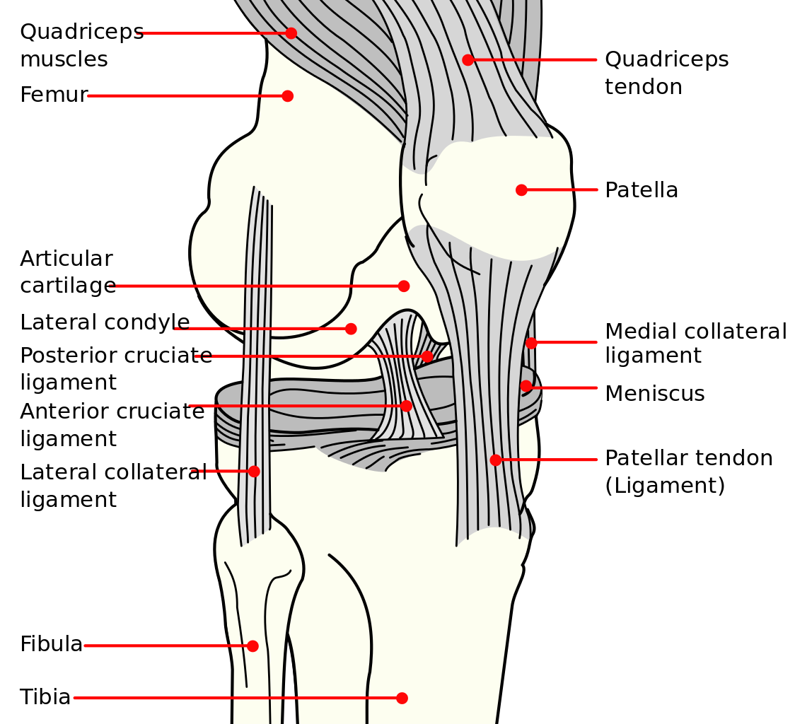 Ligaments of the Knee – Wikimedia Commons
ACL Tests
These tests assess anterior cruciate ligament integrity. The Lachman and Anterior Drawer both test one-plane anterior stability; the Pivot Shift adds a rotational component and is typically used to confirm a positive Lachman.
- Lachman Test — The most sensitive clinical test for ACL rupture (~85% sensitivity, ~94% specificity). Performed at 20–30° flexion to minimize hamstring interference. Assesses anterior tibial translation and endpoint quality.
- Anterior Drawer Test — Tests ACL integrity at 90° knee flexion. Lower sensitivity than the Lachman, especially acutely, due to increased hamstring tension at this position.
- Pivot Shift Test — Assesses anterolateral rotational instability. High specificity for ACL rupture. Used in combination with the Lachman to improve overall diagnostic accuracy.
Meniscal Tests
These tests screen for meniscal tears. No single test is highly accurate in isolation — using two or more together improves reliability.
- McMurray Test — Rotates the tibia under load to reproduce a click or pain from a meniscal tear. Medial and lateral menisci are each targeted by varying the direction of tibial rotation.
- Apley’s Test — Compression and rotation of the knee in the prone position. Pain with compression but not distraction suggests meniscal involvement rather than ligamentous pathology.
- Thessaly Test — A weight-bearing test performed at 20° flexion. The patient rotates the knee while standing on one leg, replicating loading conditions that provoke meniscal symptoms.
- Bounce Home Test — The examiner cups the heel and allows the knee to drop passively into extension. A rubbery block or springy resistance at end range suggests a meniscal tear or loose body preventing full extension.
- O’Donoghue’s Test — Applies rotation followed by valgus or varus stress to reproduce pain. Traditionally associated with the unhappy triad (ACL, MCL, medial meniscus).
PCL & Posterior Stability
These tests evaluate posterior cruciate ligament integrity and posterior tibial displacement.
- Posterior Drawer Test — The posterior equivalent of the Anterior Drawer. At 90° flexion, the examiner pushes posteriorly on the proximal tibia. Excessive posterior translation indicates PCL laxity.
- Posterior Tibial Sag (Godfrey Test) — With the hip and knee at 90°, a positive sign is visible posterior sag of the tibia under gravity — a quick screen for PCL rupture before performing the Posterior Drawer.
Collateral Ligament Tests
Valgus and varus stress tests assess medial (MCL) and lateral (LCL) collateral ligament integrity. Both are tested at 20–30° of flexion and at full extension.
- Valgus Stress Test — Applies a valgus force to the knee. Tests the medial collateral ligament (MCL). Laxity at 20–30° implicates the MCL; laxity at full extension suggests additional ACL or posterior capsule involvement.
- Varus Stress Test — Applies a varus force to test the lateral collateral ligament (LCL). Uses the same testing positions as the valgus stress test.
Rotational Instability
- Slocum’s ALRI Test — Assesses anterolateral rotatory instability. A variant of the Anterior Drawer performed with the tibia in internal rotation, isolating the lateral capsule and iliotibial band as secondary stabilizers.
IT Band
- Noble Compression Test — Screens for iliotibial band friction syndrome. The examiner applies direct pressure to the lateral femoral epicondyle while passively cycling the knee through flexion and extension. Pain at approximately 30° of flexion is a positive sign.
Patellofemoral Tests
These tests screen for patellofemoral pain syndrome (PFPS), sometimes referred to as chondromalacia patella, jumper’s knee, or runner’s knee.
- Patellar Apprehension Sign — The examiner applies a lateral glide to the patella while passively flexing the knee. Patient apprehension or guarding indicates lateral patellar instability.
- Clarke’s Sign (Patellar Grind Test) — The examiner presses down on the superior patella while the patient contracts the quadriceps. Pain or inability to complete the contraction suggests patellofemoral pathology. Note: high false positive rate — interpret with caution.
- McConnell Test — Assesses whether medial patellar taping reduces pain during a symptomatic activity (e.g., step-down). A positive response supports a patellofemoral diagnosis and may guide taping treatment.
- Passive Patellar Tilt Test — The examiner lifts the lateral patellar border with the knee near full extension. Inability to tilt the lateral edge above horizontal suggests lateral retinacular tightness.
- Waldron Test — The patient performs a slow squat while the examiner palpates the patella. Pain and crepitus throughout the movement suggests patellofemoral pathology.
- Lateral Pull Test — Observes patellar tracking during a straight-leg raise. Excessive lateral displacement (a “J sign”) suggests lateral retinacular tightness or VMO weakness.
- Zohler’s Sign — The examiner pulls the patella distally while the patient contracts the quadriceps. Pain on the posterior patellar surface suggests chondromalacia.
- Frund’s Sign — Percussion directly over the patella produces pain, suggesting patellar chondropathy.
Additional reading on orthopedic testing of the knee:

Orthopedic Assessments Made Easy: The Knee Joint Assessment
The Orthopedic Assessments Made Easy orthopedic guide book is written by Dr. Rina Pandya, a professional Doctor of Physiotherapy with more than 23 years of experience in the field, who now is a senior lecturer of Physiotherapy at the University in England after years of work in the India, USA and Oman.

Special Tests for Orthopedic Examination
First published more than 20 years ago, Special Tests for Orthopedic Examination, now in its Fourth Edition, continues to follow the authors’ initial goals of providing a simple, pocket-sized manual for practical learning purposes.
You may also be interested in…
Lists of orthopedic tests for other joints.


